Some Temperature Correlations to Hydrogel Rubber Clot in My Blood
The prior post showed hydrogel rubber clot is suppressed by a normal body temp of 100°F/38°C. Let's refine the details.
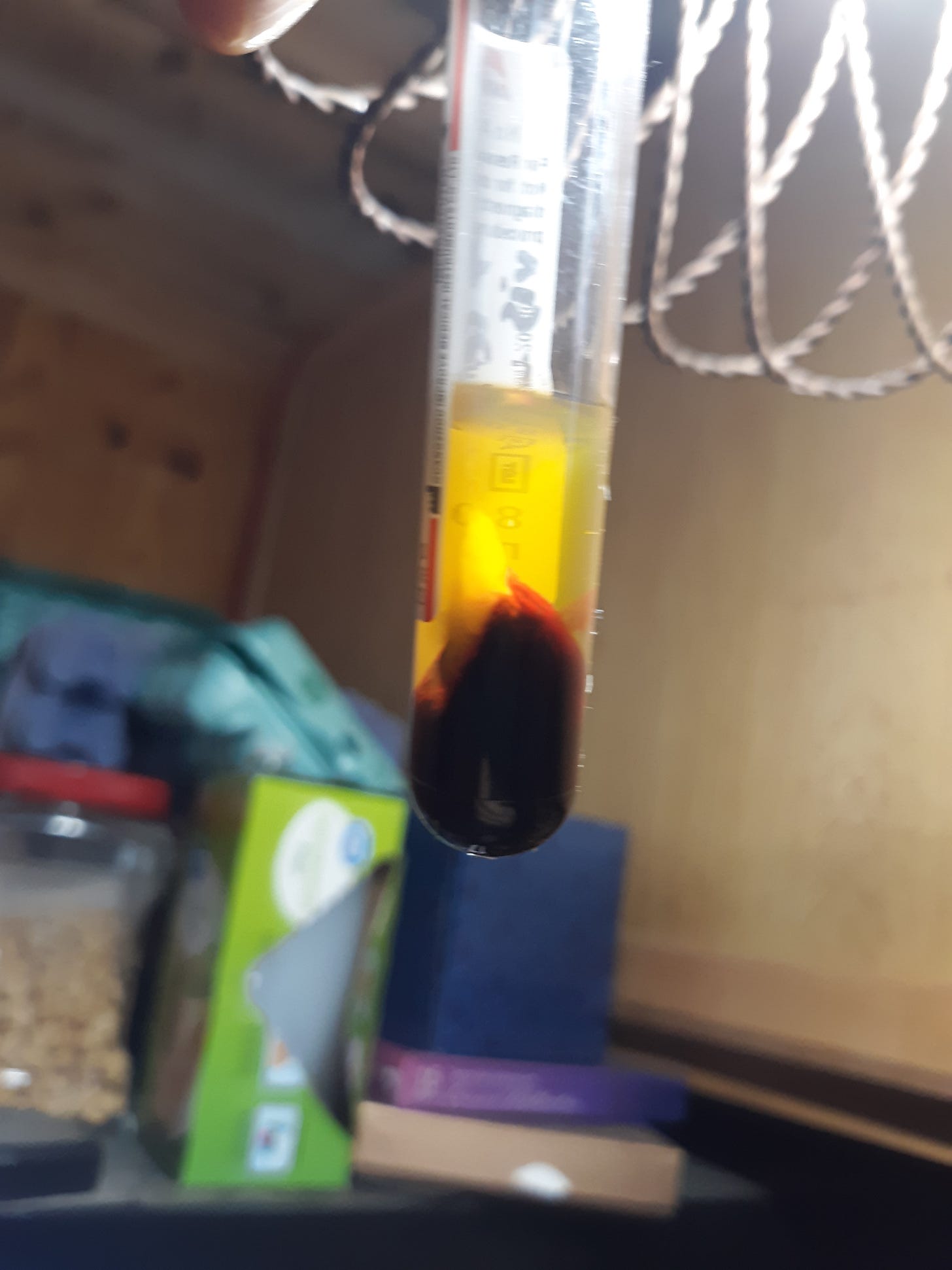
The control here is to collect blood known to contain hydrogel rubber clot (myself) into a plain vacutainer tube at room temp of about 75°F/ 24°C and allow normal blood clotting to occur for 30 min in the tube, then centrifuge for 30 min at room temp, then visually examine tube for hydrogel rubber clot. The following pic shows a cloudy area above the packed red cells consistent with hydrogel rubber clot.
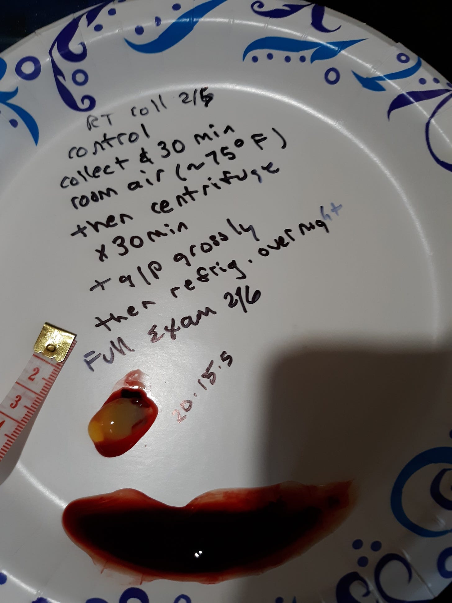
The tube is then refrigerated at 42°F/ 5.5°C overnight and full exam done by emptying tube contents. Hydrogel rubber clot is noted with initial dimensions of 20x15x5 mm.
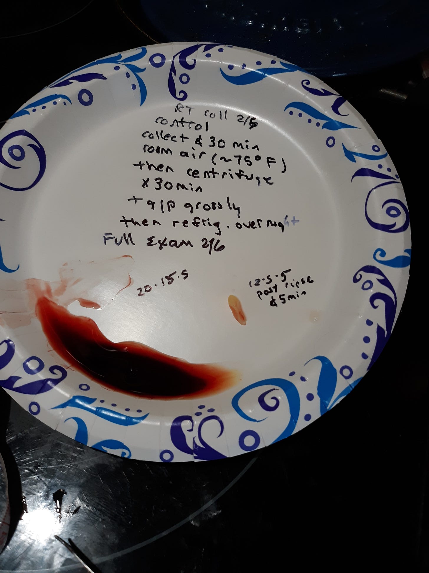
If the hydrogel rubber clot is rinsed in tap water it shrinks and becomes whiter in about 5 min taking on the rubbery calamari state as the next pic shows.
Next I collected my blood into a plain vacutainer tube and immediately placed as soon as practically possible into an incubator at about body temp of 100°F/ 38°C for the initial 30 min during which normal blood clotting would occur in a test tube. The tube was then centrifuged x 30 min at room temp of 75°F/ 24°C and then visually inspected for hydrogel rubber clot. The next pic shows these results and noted a possible small amount of hydrogel rubber clot.
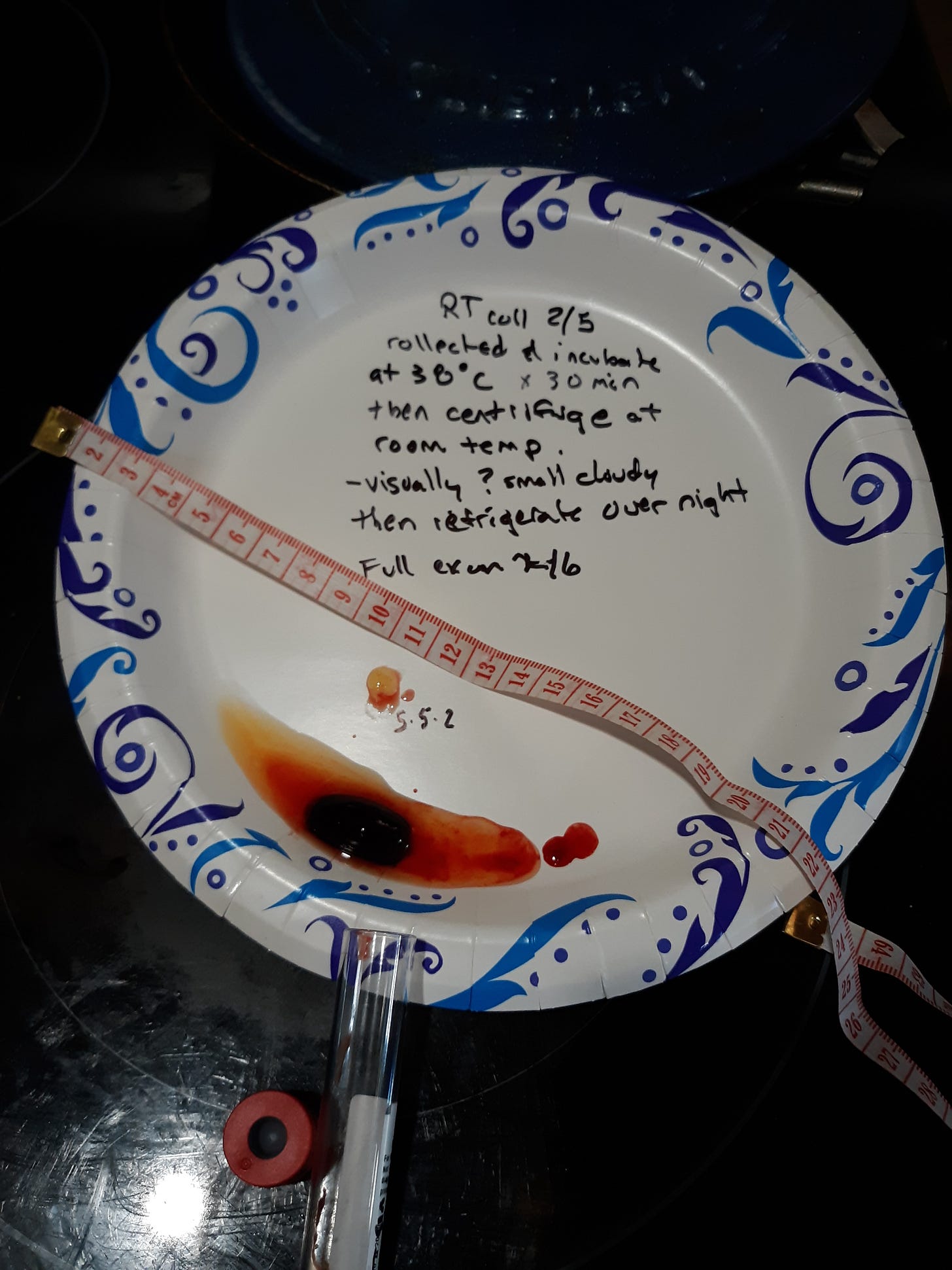
Full exam was performed the next day after refrigeration overnight at 42°F/ 5.5°C. The results are seen in next pic with hydrogel rubber clot reduced 50 x compared to control.
After rinsing and given 5 min time the hydrogel contracts and becomes whiter and rubbery as shown.
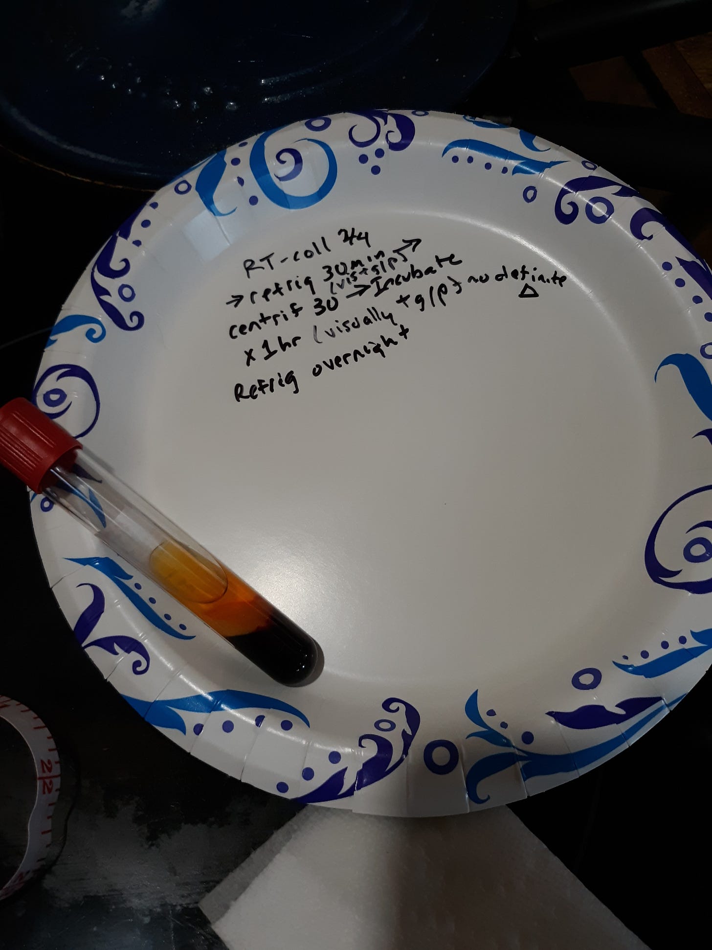
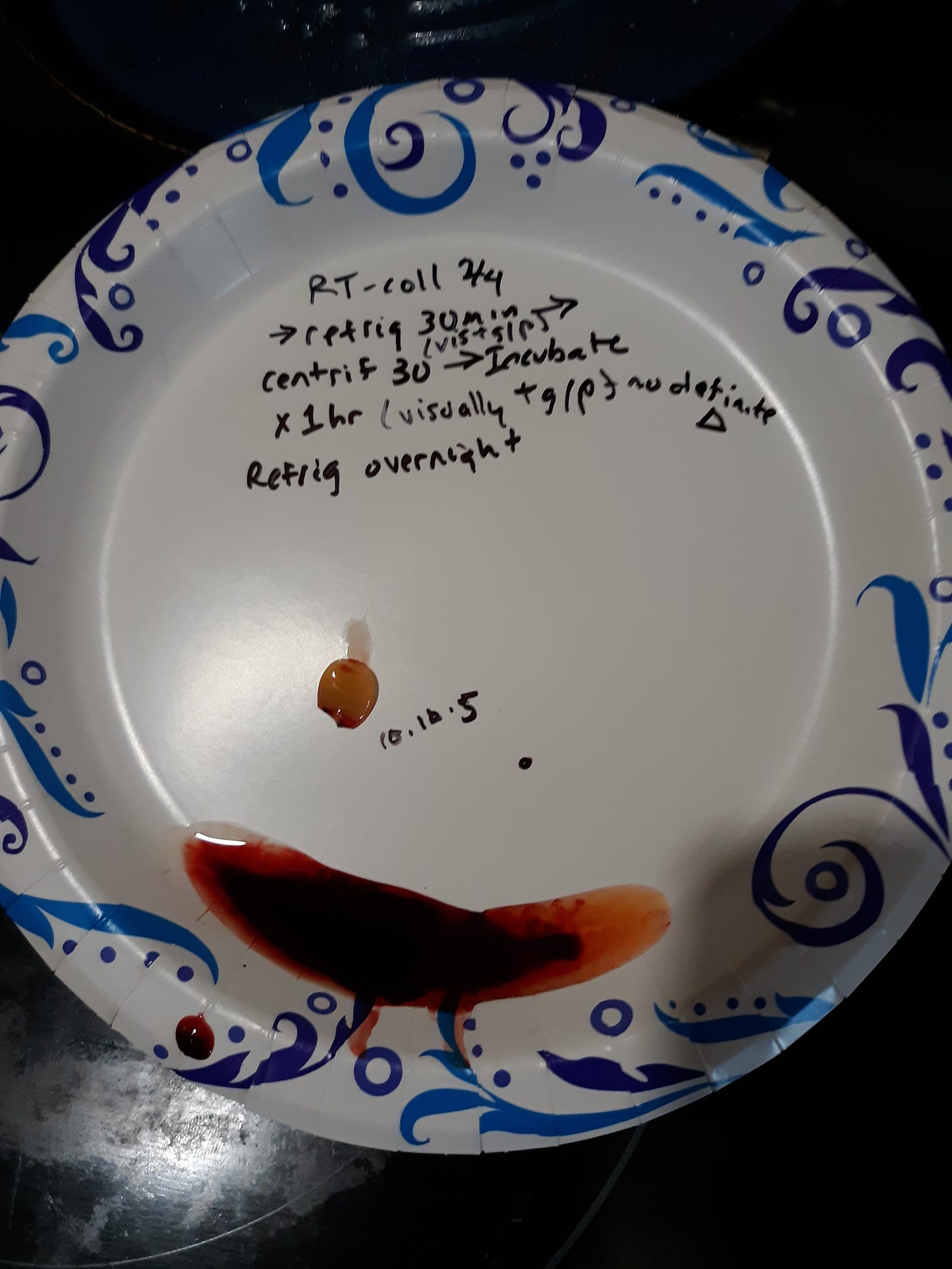
In the next several experiments I wanted to refrigerate the samples during the initial 30 min after blood collection. This initial 30 min is when normal clotting will occur in a test tube. The control here is a plain vacutainer tube filled with blood and placed in refrigerator at 42 °F/ 5.5°C for 30 min then centrifuged in room air 75°F / 24°C for 30 min. I expected to see hydrogel rubber clot and this was observed as the pic shows. I also wanted to see if the hydrogel rubber clot would re-dissolve if the tube was heated to body temp for the next hour. No obvious decrease in hydrogel size was noted after the hour in the incubator. Following this the tube was refrigerated overnight.
Full exam is shown.
A variation of the experiment is as follows. Take the sample of blood and refrigerate at 45°F /5.5°C for 30 min after collection then incubate the specimen at body temp for an hour followed by centrifugation for 30 min at room temp. The next pic shows that no hydrogel rubber clot is seen.
Full exam was performed the next day after refrigeration overnight. A tiny fragment was isolated but did not appear to be hydrogel rubber clot. It disintegrated when rinsed.
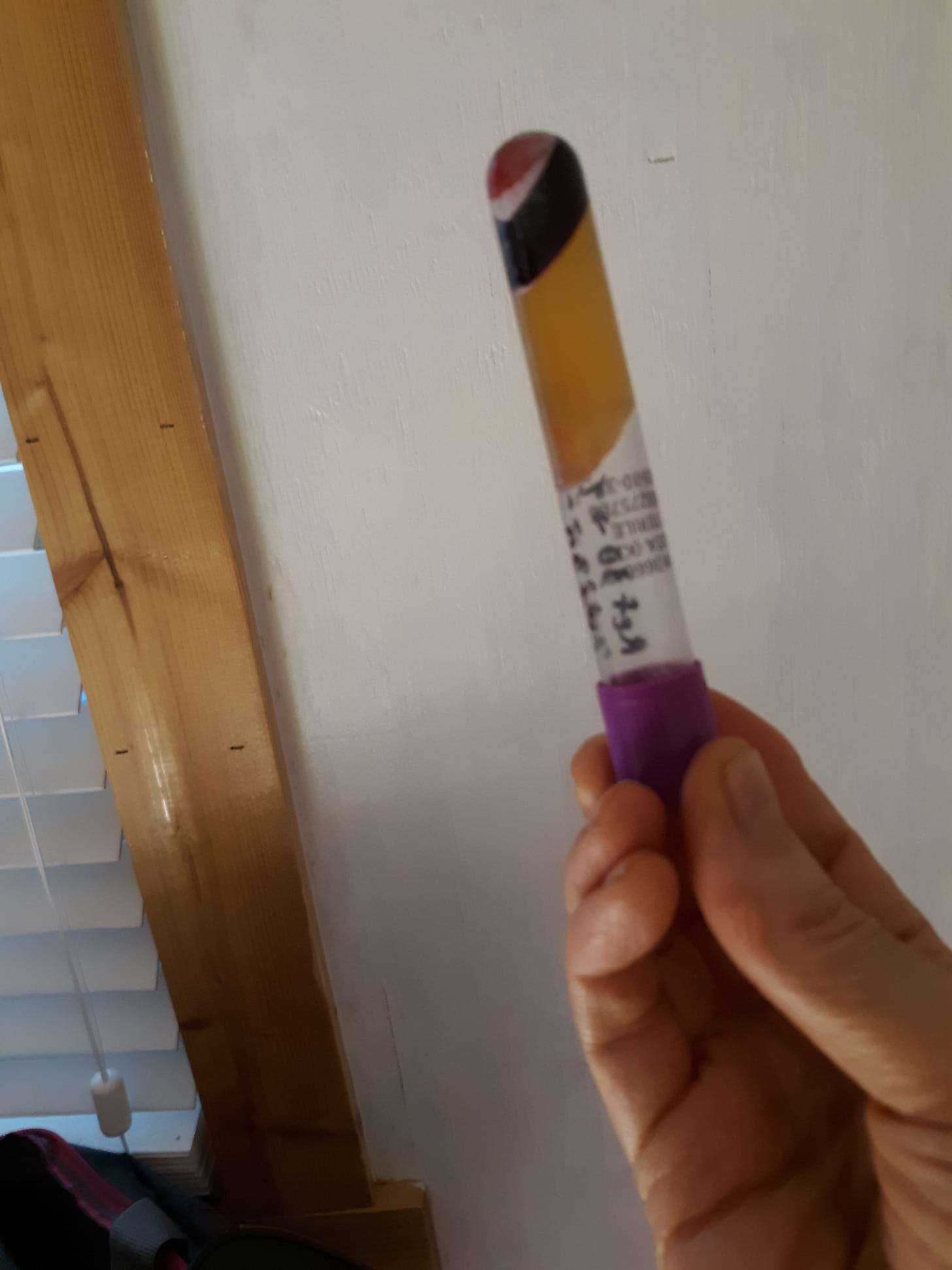
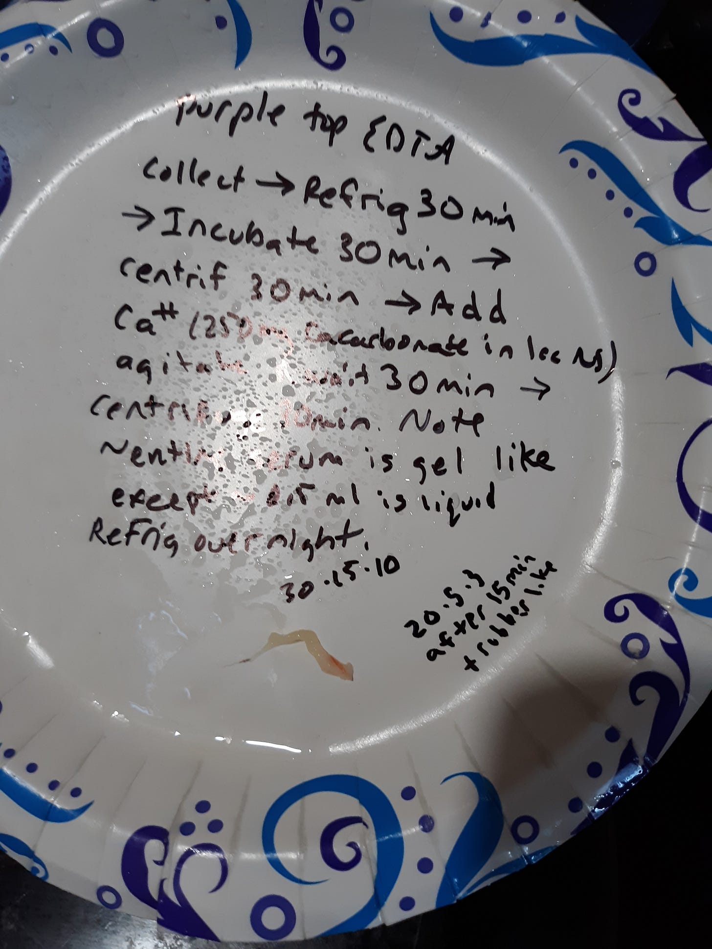
A further experiment consisted of filling an EDTA vacutainer tube with blood and refrigerating the sample for the initial 30 min following collection. EDTA is an anticoagulant. The tube was then incubated at body temp for 30 min followed by centrifugation for 30 min. I did not expect to see hydrogel rubber clot since I had shown hydrogel rubber clot will not be expressed if normal clotting is inhibited by anticoagulants such as EDTA or citrate or heparin. I next neutralized the anticoagulant effect by adding 250 mg calcium carbonate in 1 ml normal saline to the blood tube and allowed to mix for 30 min. I then centrifuged the tube for 30 min. I noted nearly the entire serum area to be a gel as the following pic shows.
I refrigerated the tube overnight for full exam the next day as seen in next pic.
The gel was rinsed and in about 10 min the gel became whiter and rubbery as the next pic shows.
Summary and Discussion. The hydrogel rubber clot is suppressed by a normal body temp. It was shown that hydrogel rubber clot was 50 x less when maintained at body temp during the first 30 min when normal clotting takes place in vitro compared to what was seen in the control collected at room temp and maintained at room temp during this initial 30 min. I think if it was possible to maintain the entire blood collection and initial 30 min during which clotting occurs at body temp then complete suppression of hydrogel rubber clot would be seen. I know this summer I drew blood on two individuals when outside temp was near 100°F and took the specimens home in a hot car and results were negative for hydrogel rubber clot. I suspect these were false negatives.
To summarize, in order for hydrogel rubber clot to be expressed in an individual known to have it, blood collected needs to clot during the first 30 min following collection at a temp less than normal body temp. The needed lower temp value is unknown but 75°F / 24°C was sufficient in this testing.
Please note in the summary that the normal blood clotting pathway needs to be intact and if full anticoagulation is present using EDTA, citrate, or heparin then hydrogel rubber clot will not be expressed. (I had shown this in a prior post).
If I had to offer a possible explanation to cover these findings it would include a theory that something is released during normal blood clotting that acts on the precursors to hydrogel allowing them to entangle with each other and capture water forming a 3D gel. If you are a believer in spike then certain proteases can denature spike and allow it to form beta pleated sheets that can form an amyloid hydrogel. Remember that during the normal blood clotting cascade many specific proteases are activated and could these be acting on hydrogel rubber clot precursors? Could the precursors have a temperature sensitive range in order to be denatured and take a form that can entangle with each other forming a hydrogel. Here is a paper that speaks of amyloid and hydrogels as well as protease.
The hydrogel rubber clot behaves as a themo-sensitive hydrogel. The following paper describes some of these.
Section 2.2 on Positively Sensitive Hydrogels reports the following. Hydrogels are believed to have a random coiled conformation at higher temperatures, while they begin to form a double helix and an aggregate with the decrease of temperature, which act as a physical junction of gel. Owning a large number of reactive functional groups gift the gelatin ability of modification. Yang and Kao (2006) synthesized a hydrogel composed with gelatin and poly(ethylene glycol)-poly(D,L-lactide)(mPEG-DLLA) block copolymers, which could flow at 37 °C and gel at room temperature.
This might relate more to you all. Gelatin, like Jello, is a temp sensitive reversable hydrogel made up of denatured collagen. Remember the late German pathologist, Arne Burkhardt, felt the hydrogel is the result of blood vessel destruction caused by an autoimmune reaction to spike proteins being produced in vessel tissues so denatured collagen should be a component of the hydrogel rubber clot according to him. I did a few experiments here at home with gelatin and noted its reversibility from gel to solution with temp changes. In the above set of experiments I did not see any reversibility.
To review from the prior post. 1. The collected blood must not be fully anti coagulated with EDTA, citrate, or heparin. In a fully anti coagulated state with these present any hydrogel rubber clot will be prevented. The same prevention can occur with high dose vitamin C. It should be noted that adding back calcium to the blood in EDTA or citrate containing tubes can cause the hydrogel rubber clot to be expressed.
2. Body temperature appears to prevent the expression of hydrogel rubber clot.
3. Hemolysis (rupture of red blood cells) prevents the expression of hydrogel rubber clot.
Praise the Lord. Praise, O servants of the Lord, Praise the name of the Lord! Psm 113 :1







Very interesting experiment Ron. I believe flow rate and temperature are large cause, also photoswitchability. The hydrogel is also tunable by photoswitchability. What I find even more interesting is calcium causes formation.
Photoswitchability also causes the hydrogel to lose tunability over time, or the amount of times it's photoswitched. I believe a Ca2+ is exchanged for a nanoheavy metal such as Zn2+, Mn2+, Ect. So the Ca2+ are the more clear glass like layer. The yellow/white layers is the slightly still photoswitchable layers so a mixture of calcium and nanoheavy metals.
Something like that anyway. People dying while swimming in cold water. Athletes who's heart rates and blood pressure drops phenomenally low when in deep sleep, then die when playing at the peak of their sport. Starts to make more sense hey?
Remote assassination... Via EMF is another kettle of fish but similar. They can use nano bio drones. I experienced it. Had the white clots grow in some veins, clotting is how it starts.... Just got rid of Lyme's disease too.
This is how I stumbled upon the photoswitchability using green laser, and a weak chelator Copper(ii) glycinate, along with the rest of the protocol I was taking. That disassembled the white clots.
I'll write about it soon. ☺️
My docs are not keen on pain control and my old stand by is aspirin. W/Von Willebrand's and almost daily aspirin I am clotting for the first time at age 64. No surprise, I still bleed for several hours but now there is clotting.
I did not participate in the C-19 vaccine campaign.
I suspect my exposure to the spike protein was Factor 8 for an emergent procedure. (I respond poorly to DDAVP.)
Myocarditis 3 months after infusion. Now I take IVM every few weeks, as that is how long it takes to get gummed up w/fragmented ecg's, brain fog... I suspect that whole blood products are able transporters.