
Out of Remission. Time for a new study. Rubber Clot in My Blood.
Vacutainer tube blood collection and use of a centrifuge is a good way to detect the presence of hydrogel, rubber clot in the blood.

Over the past month I have made a few changes in my treatment for the hydrogel, rubber clot in my blood but I have monitored the effect by vacutainer tube blood collections.
Let me review the technique since I believe it is the gold standard to verify effectiveness of any treatment for the hydrogel, rubber clot, in a living person’s blood. Compared to live blood analysis, there is less interpretation needed to verify effectiveness. The hydrogel rubber clot is either present or absent by the test.
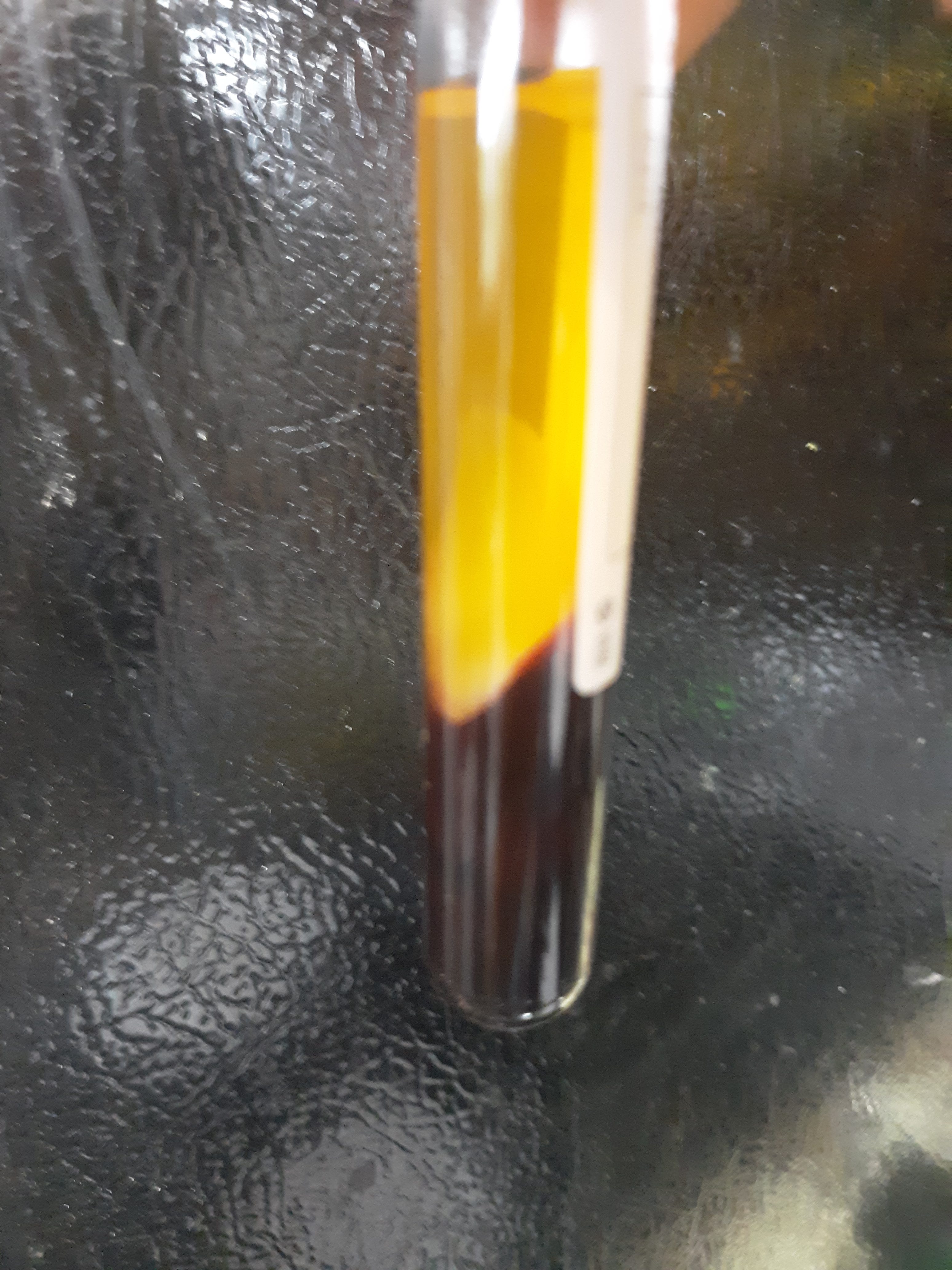
Blood is collected by venipuncture into a plain (not contain anti coagulant) red top vaccutainer tube, the tube gently inverted ×10, allowed to coagulate for 30 min, the tube then centrifuged x 30 min, a visual inspection of the area above the packed red cells can be done to look for any cloudiness that is likely hydrogel rubber clot, tube is placed upright in refrigerator overnight, and final inspection is done next day by emptying the tube contents on a paper plate to visualize the hydrogel rubber clot if present.
Here is a recent example. The cloudy area is seen above the red cell clot is hydrogel rubber clot. The fluid is of course the straw colored serum.
This shows examination and isolation of hydrogel rubber clot from main clot with forceps (forceps not shown).
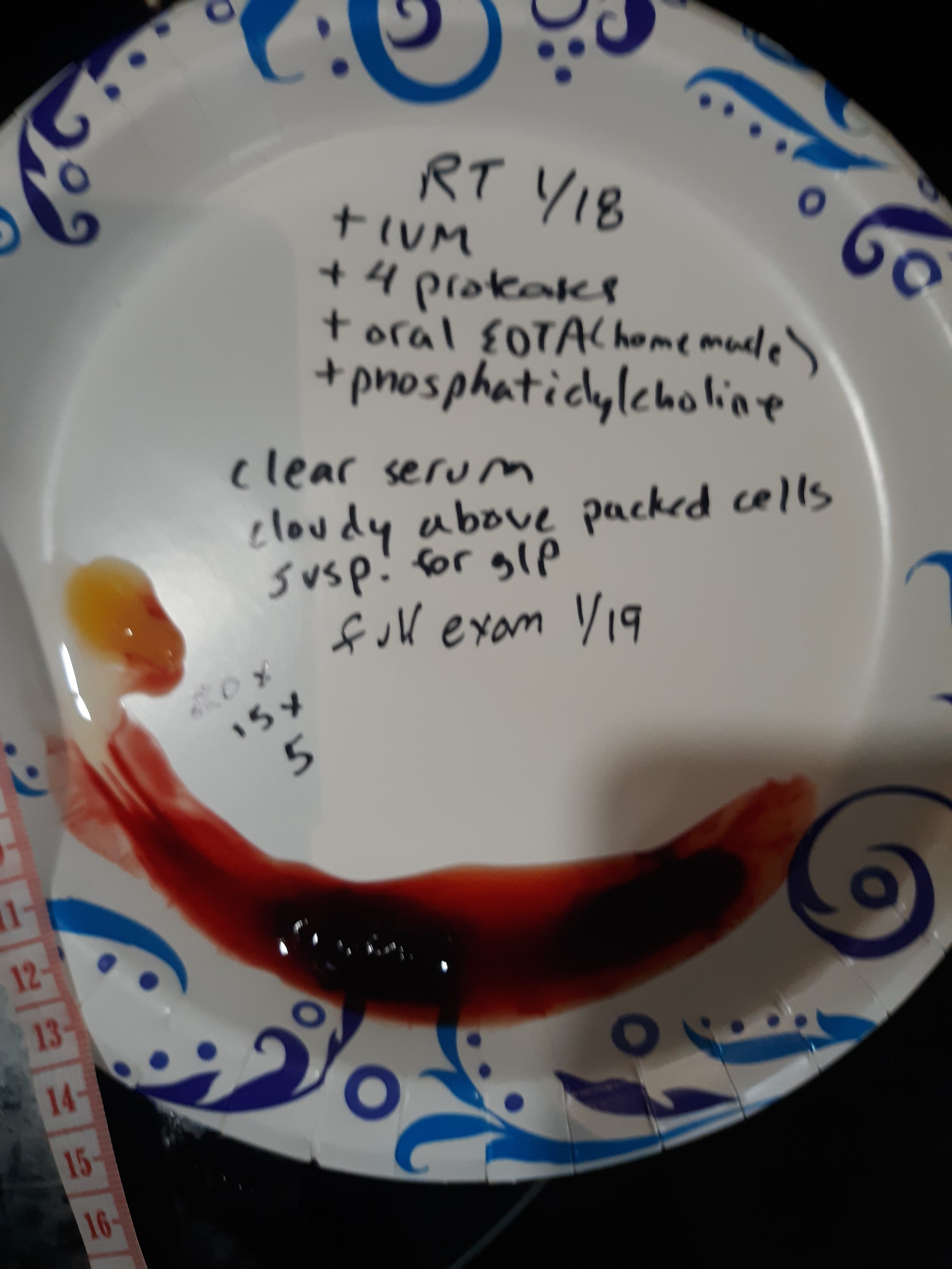
This slide shows the hydrogel rubber clot about 5 min after isolation and rinsing in tap water. It becomes more firm and white as well as a rubbery consistency (like calamari). I referred to the hydrogel as g/p (gel/plastic) in the past and this notation is on the plate as well as the hydrogel measurements in mm before and after rinsing plus 5 min.
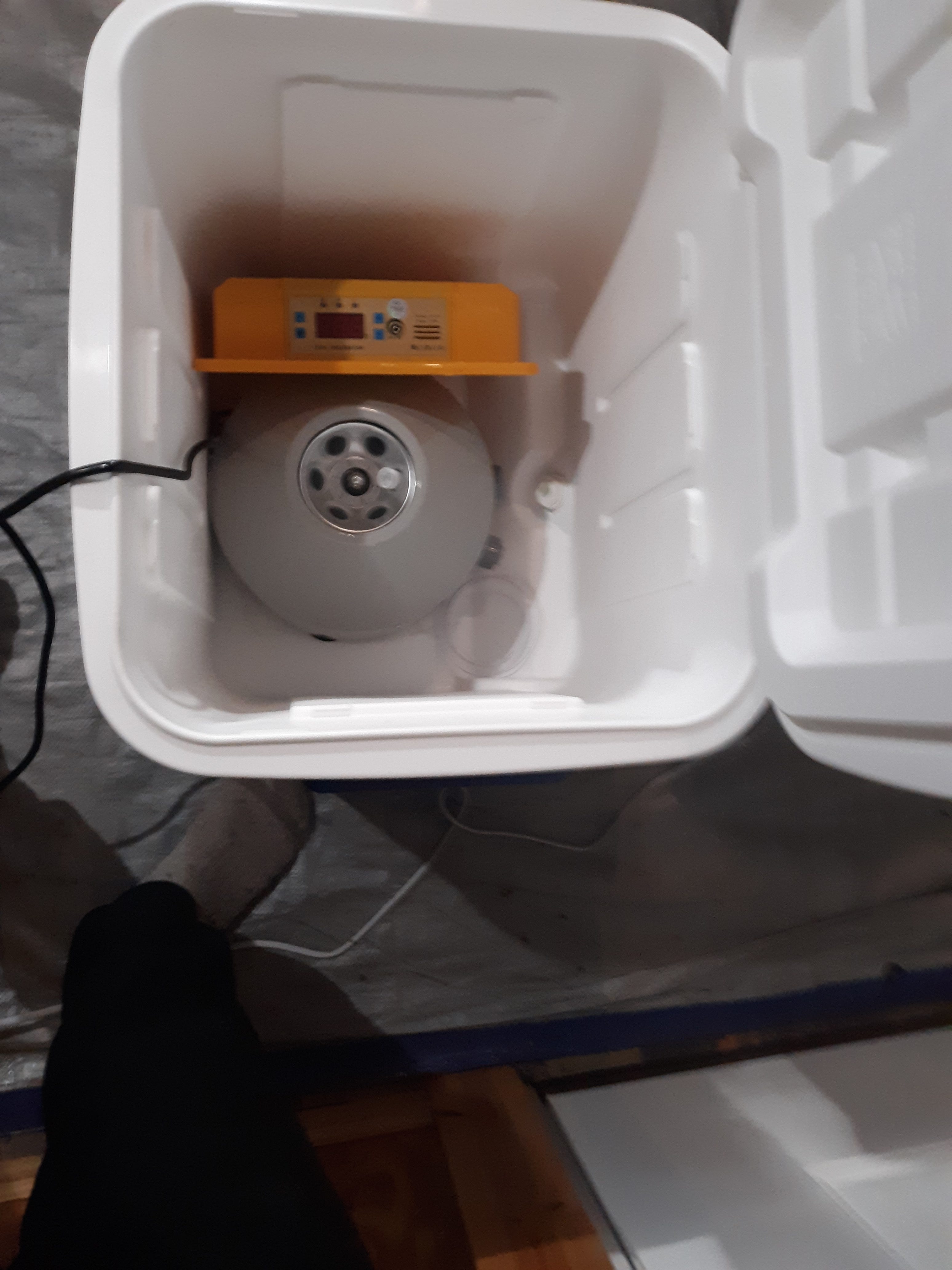
Most recently I made some changes in my treatment regimen. I continued daily oral ivermectin at 20 mg per day. I weigh 140 lbs so this dose is 0.3 mg/kg. Remember during the pandemic the initial dose was 0.2 mg/ kg but increased to 0.6mg/kg during the delta strain. I personally use the liquid ivm that recommends 1 ml (10 mg) per 110 lbs cattle weight. I continued the 4 different oral proteases throughout the day by taking one type half hour prior to a meal or 2 hrs after a meal. The proteases are nattokinase 2K FU, lumbrokinase 150 mg, serrapeptase 240 K SPU, bromelain 500mg. I add curcumin 500 mg, and NAC 1000 mg (augmented best). I also was taking a EDTA supplement like MedFive each day. The manufacturer of the MedFive product had some production issues and said was on back order so when I ran out I substituted an oral liposomal EDTA that I had mixed on my own but was 400 mg per dose. I also added a phosphatidylcholine commercial product. The hydrogel rubber clot returned and the above pictures show this. This is actually the result I showed in a past post indicating the MedFive product was helpful. Since I was out of remission I wanted to test the theory that the appearance of hydrogel rubber clot was dependent on a drop in temperature of the collected blood. This idea is supported by the observation that people known to have hydrogel (like myself) are not manifesting rubber clot formation and its issues during everyday life (usually). So I need to collect the blood and place the tube of blood in an environment at body temp for the 30 min time given to clot and then centrifuge at body temperature for 30 min per routine and keep at body temp overnight. I did this by using a large insulated ice chest that could hold my centrifuge and the heated top unit of my chick incubator that has a digital thermometer.
Here is a picture of the set up.
I collected the blood known to contain hydrogel plastic clot by the control sample taken the same day but collected a second specimen kept at approximately body temp of 38 degrees C (100.4 farenheit). A quick look showed the serum was straw colored with no evidence of hydrogel rubber clot after centrifuge process at body temp but needed to be also kept at body temp overnight. The specimen was kept in the incubator overnight at 38 C and visual inspection of the tube was negative for hydrogel rubber clot as noted by the non- cloudy serum above the packed cells as the following picture shows.
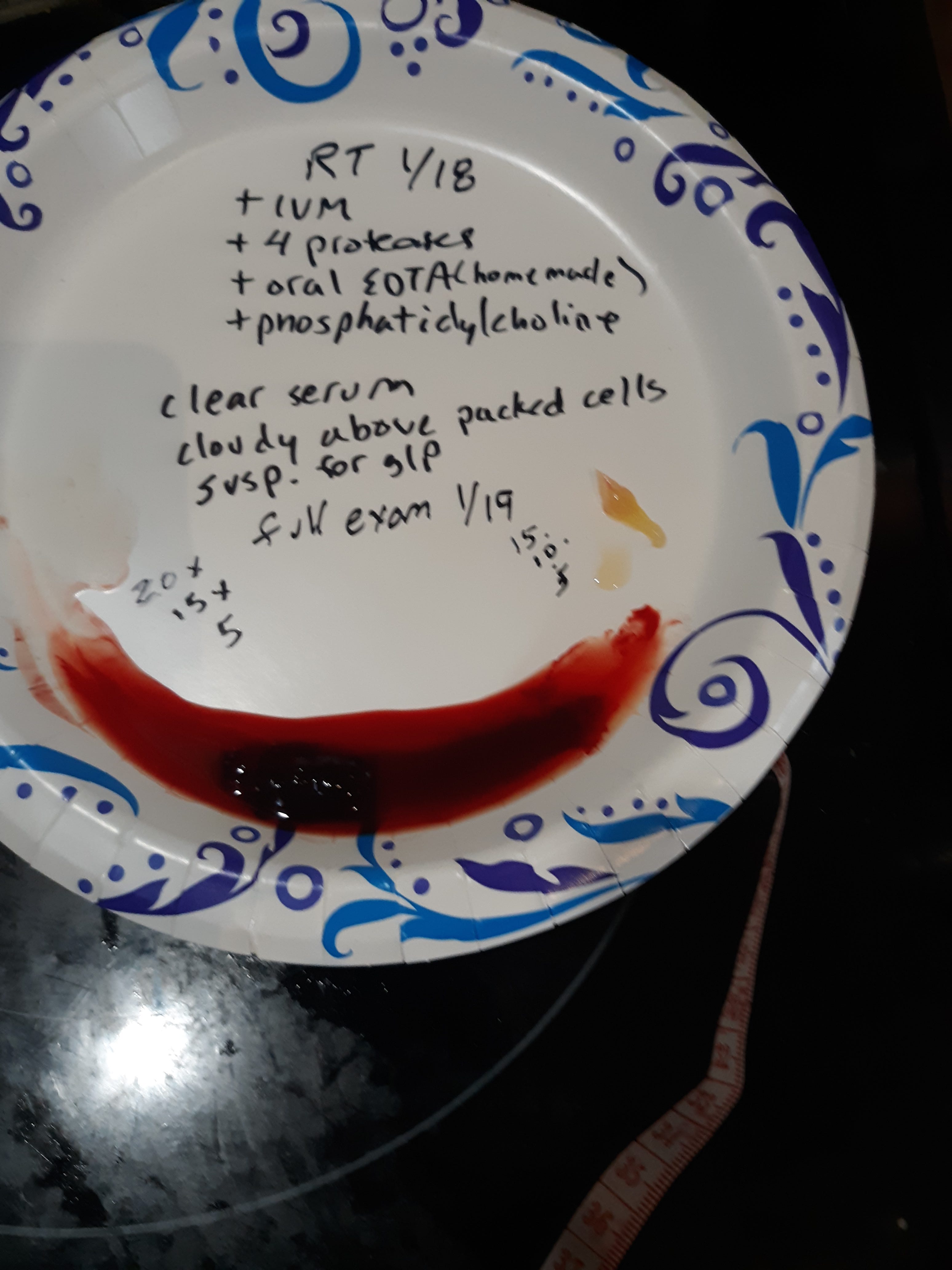
I then kept the tube at room temp for half an hour and then refrigerated the tube for 30 min. I then examined the tube further by shifting the contents and noted what appeared to be the normal currant jelly clot to be present but again no hydrogel rubber clot was present. I then centrifuged the tube at normal room temp for 30 min. No obvious hydrogel rubber clot was present in the tube that was kept at body temp for the initial processing and overnight. I then refrigerated the tube for 4 hours. Final exam was done by emptying the tube contents and no definite hydrogel rubber clot was seen as next picture shows.
Summary and Discussion. I again refer to the late German pathologist, Arne Burkhardt, that demonstrated the hydrogel plastic clot from a living person in a technique similar as demonstrated here using plain vacutainer tubes, a centrifuge, as well as refrigeration. He mentioned that the hydrogel rubber clot was expressed with aid of refrigeration. Dr Burkhardt felt the hydrogel rubber clot was a mixture of spike, proteins from the autoimmune destruction of the blood vessel wall due to the production of the foreign spike protein in the infected vessel tissues, and amyloid. Amyloid may result from a denatured protein that demonstrates a beta pleated sheet configuration such as when spike is acted upon by neutrophil elastase. Here is the link to Dr Burkhardt on rumble: https://rumble.com/v2h5zuy-pathologist-dr.-arne-burkhardt-autopsies-show-the-mrna-vaccine-shreds-peopl.html . This is a reference to how spike can form amyloid segments.
I theorize that normal blood clotting releases other proteases that denature other proteins present that can form hydrogel.
I have shown and posted the following are required to have hydrogel rubber clot expressed in vitro in an individual known to have it present in their blood.
The collected blood must not be fully anti coagulated with EDTA, citrate, or heparin. In a fully anti coagulated state with these present any hydrogel rubber clot will be prevented. The same prevention can occur with high dose vitamin C. It should be noted that adding back calcium to the blood in EDTA or citrate containing tubes can cause the hydrogel rubber clot to be expressed.
Body temperature appears to prevent the expression of hydrogel rubber clot.
Hemolysis (rupture of red blood cells) prevents the expression of hydrogel rubber clot.
These are some findings I noted with testing. These findings are not mutually exclusive and all occur at the same time. I say this because blood banks would be seeing hydrogel rubber clots all the time if only lower temperature was needed to express hydrogel rubber clot. I have heard of the rubber clots appearing in plastic tubing during some blood removal processes. Last year the bodybuilder Jo Linder spoke of white clots seen during his plasmapheresis (removal of plasma) treatments as seen at 4:30 of this video. It should be noted that blood banks usually use acid citrate dextrose as an anticoagulant. https://rumble.com/v2y7p48-fitness-guru-bodybuilder-jo-lindner-30-died-w-aneurysm-after-4-covid-vax-7..html
In the video with Dr Arne Burkhardt he notes frequent aneurysms of vessels due to the autoimmune destruction of tissue. I recall Dr Ana showing a picture of hydrogel rubber clot in tubing sent to her during some extracorporeal blood treatment.
In summary, it is possible to prevent the expression of hydrogel rubber clot in vivo using several compounds. This is my experience and not a universal recommendation although very similar to protocols recommend by Dr Peter Mccullough and others. It includes ivermectin, proteases, (nattokinase, bromalin, serrapeptase, lumbrokinase), curcumin, NAC, and the MedFive EDTA containing product. Dr Ana Mihalcea is an advocate for EDTA. Again we need others doing similar testing and sharing their results.
The Lord also will be a refuge for the oppressed, A refuge in times of trouble. Psm 9: 9
Thank you for all your work. If I was employed I would definitely subscribe to yours and others Substack research posts. I do tweet your work, and put them in my Bioweapons Facebook post, and I have two Substack posts dedicated to the biowarfare, but I've taken a broader view. Cheers, and thanks again.
You might also try (1) BORAX, which is widely thought to stall nano growth. Start with 1/8 tsp twice a day and work up to 1/2 tsp twice a day. (2) Methylene Blue, which Dr. Ana says is a go-to for dissolving hydrogel. Dr. Levy, in his recent interview with her, showed that he was quite enthusiastic about MB.