Is the Platlet Rich Fibrin (PRF) Rubber Clot the Same as Dr Ana's Syringe Clot?
Most of us follow the work of Dr Ana Mihalcea and have seen her show rubber clots from living patients formed in a large syringe from blood obtained from her patients.
The PRF method is performed by collecting blood via venipuncture, immediate centrifuge for 8 min at 1,500 rpm, isolating the fibrin rich layer, and squeezing out excess serum to show a white and rubbery clot (calamari effect).
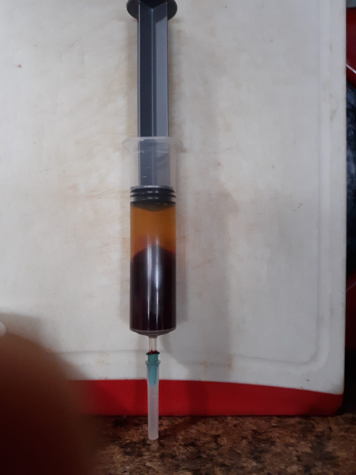
Is this PRF rubbery clot the same as the rubbery clot seen in the large syringe of Dr Ana? Follow her at anamihalceamdphd.substack.com. That clot is formed from 30 ml blood drawn via venipuncture into a large plain plastic syringe and allowed to set for 4 hrs or overnight at room temperature.
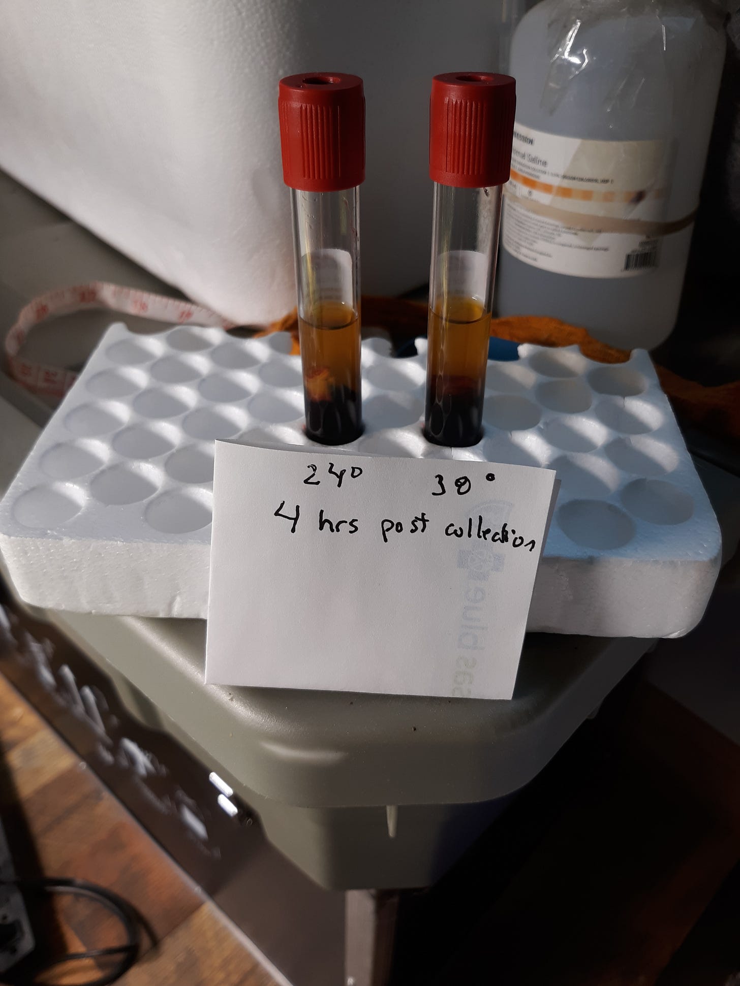
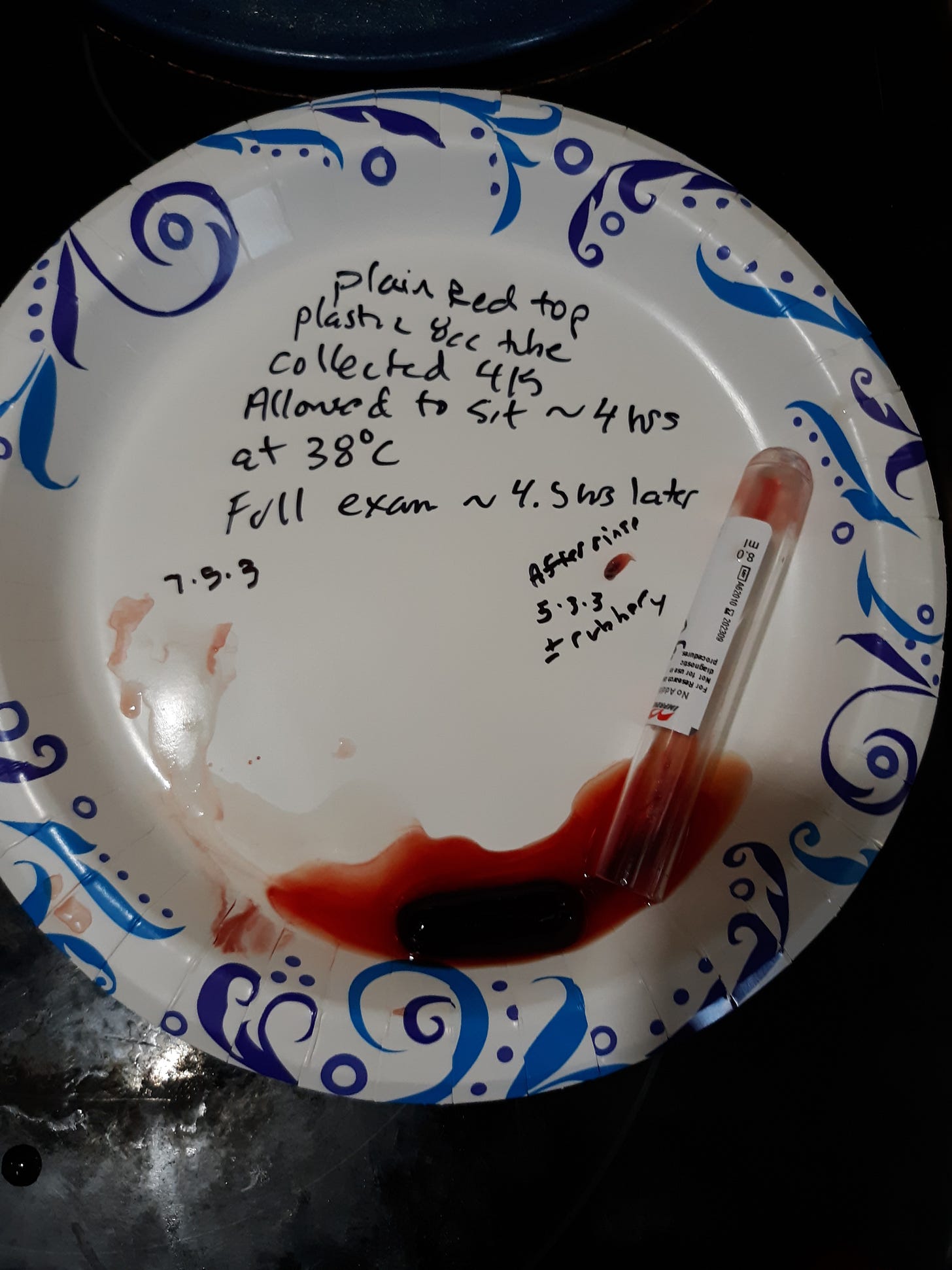
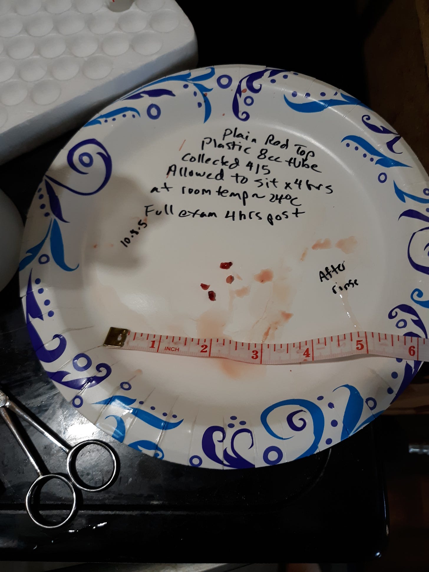
A variation to the syringe of blood would be a 8 to 10 ml plain plastic vacutainer tube. I drew two 8 ml plastic red top vacutainer tubes with blood. One tube I allowed to sit for 4 hrs at room temp of 24°C(75°F) and the other I placed in an insulated ice cooler at 38°C(100°F) for 4 hrs. Here are the tubes after 4 hrs.
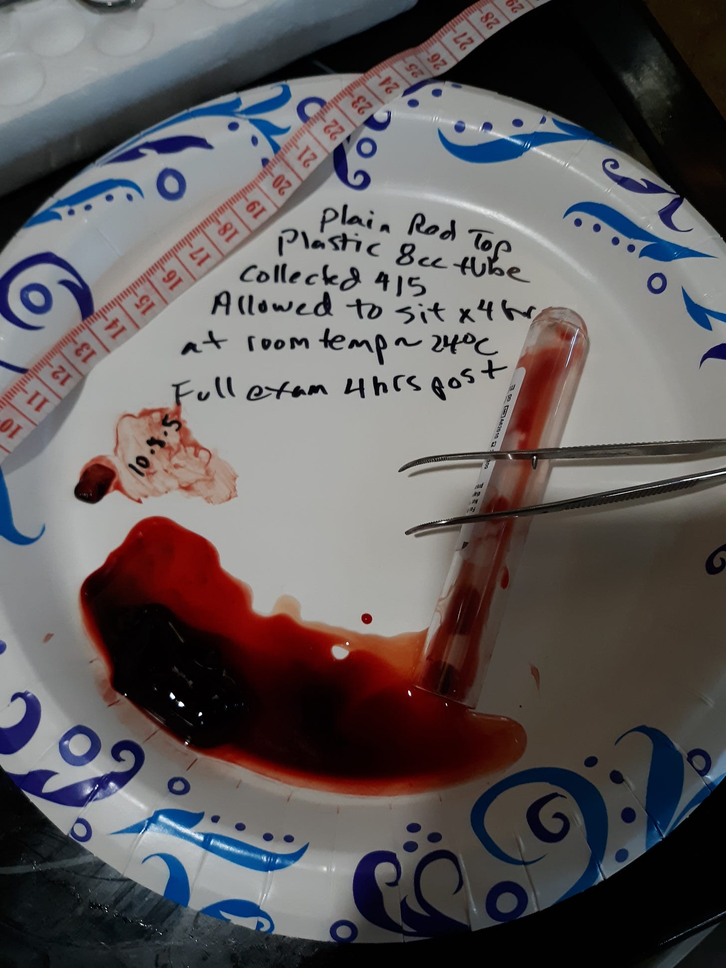
There is obvious presumed rubber clot in the tube kept at room temp and something less in the tube kept at 38°C(100°F). I performed an exam on the tube kept at room temp as the following picture shows.
The rubber clot was rinsed and it appeared more white and rubbery but did not shrink much of notice.
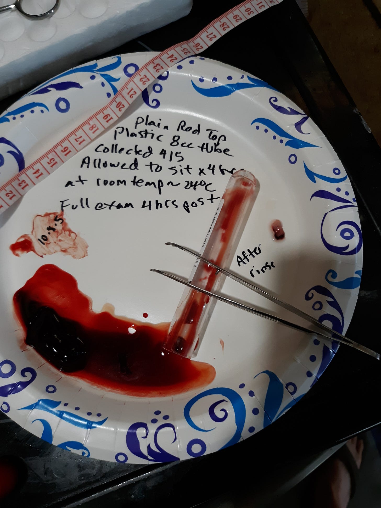
I examined the tube contents kept at 38°C(100°F) for 4 hrs. The area of interest showed the following.
After a rinse in tap water the possible rubber clot was noted not to be very rubbery and broke up a bit. I will say at this time I will need to repeat this attempt to see if this area demonstrates a temperature sensitive hydrogel.
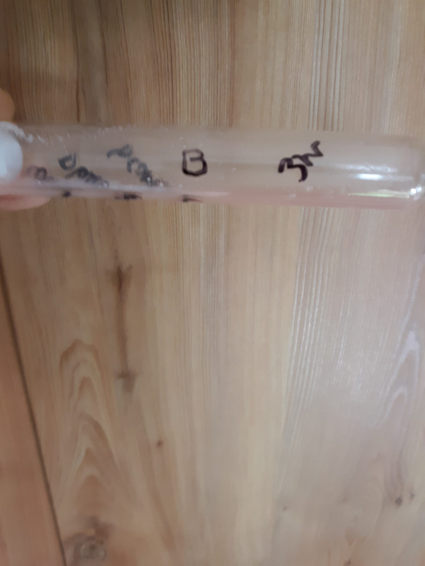
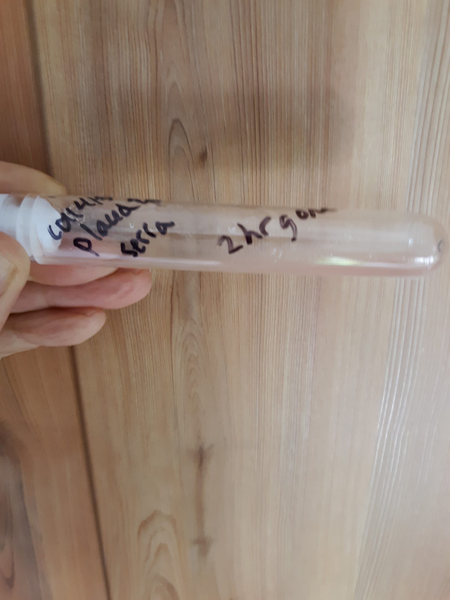
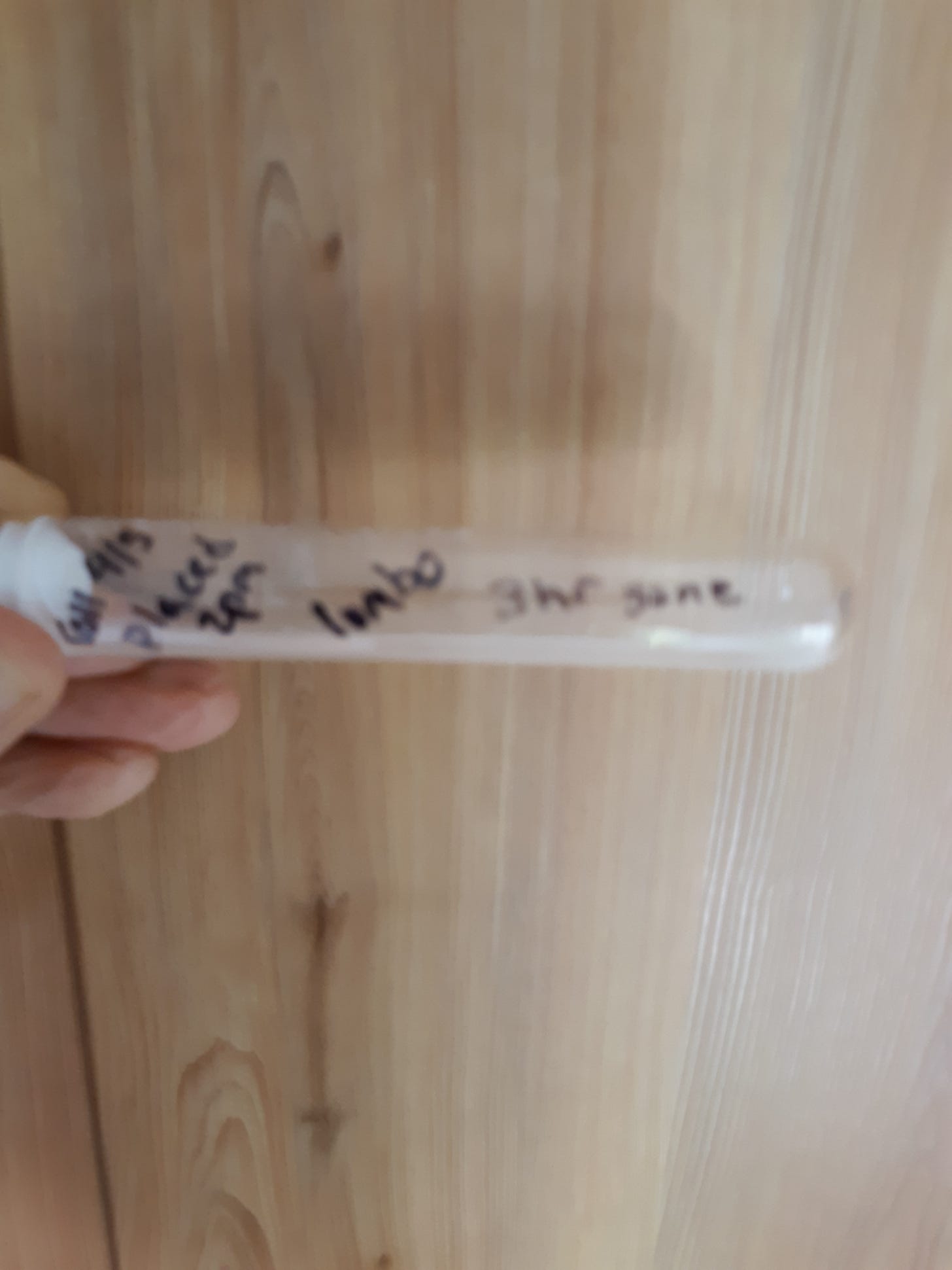
I next wanted to see if rubber clot obtained at room temperature was able to be broken down by fibrin proteases so I cut it into 4 pieces of about 5 x 2 mm and placed in vials containing about 0.05 mg of either bromelain, nattokinase, serrapeptase, or lumbrokinase and a few ml of normal saline. The following pic shows the specimens.
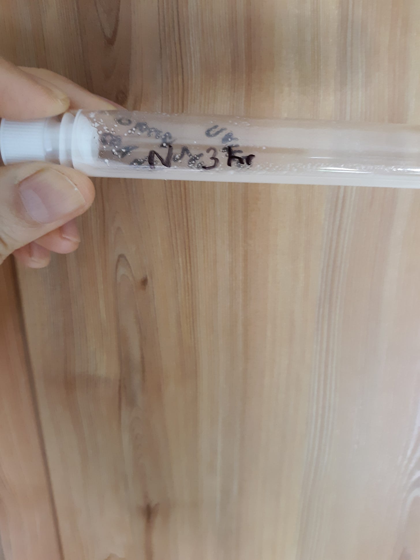
I placed a fragment of rubber clot into the various proteases and the fragments were dissolved as the following pics indicate. Bromelain dissolved it within 3 hrs.
Nattokinase dissolved it within 3 hrs.
Serrapeptase dissolved it within 2 hours.
Lumbrokinase dissolved the fragment within 3 hrs.
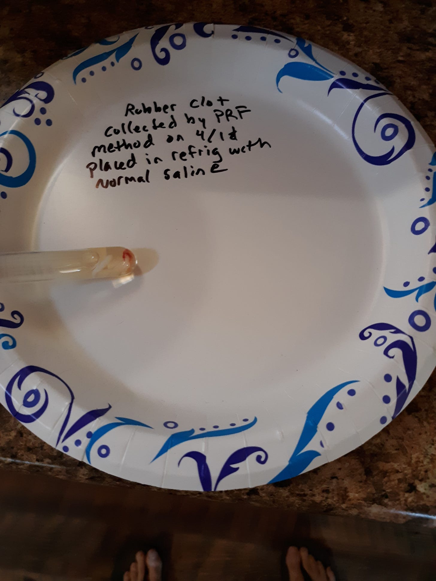
In my prior post I generated rubber clot via the PRF method and could not show evidence of a temperature sensitive nature to the fibrin rich clot. I took the PRF rubber clot from 4/1 and cut it up in similar fragments to place into the above tested proteases. Here is a fragment in a tube prior to being dissolved so you can have an idea what it looked like. I cut the rubber clot into 5×2 mm fragments to place into protease containing tubes.
The bromelain dissolved the clot in 3 hrs.
The nattokinase dissolved the clot within 3 hrs.
The serrapeptase dissolved the clot within 3 hrs.
The lumbrokinase dissolved the clot within 3 hrs.
Summary and discussion. I would answer the question that I am not sure if the PRF rubber clot is the same as the rubber clot obtained by allowing blood to sit for 4 hrs in a tube. My suspicion is that they are not the same. Obviously the PRF rubber clot is rich in fibrin and the 4 hr rubber clot is at least much less in fibrin by the fact that it is above the currant jelly clot known to be held together by fibrin.
The good news is that both variety of rubber clot can be dissolved by the four fibrin proteases tested.
Other differences is the PRF method shows a fibrin rich clot that remains an obvious hydrogel in the serum and can be squeezed out to get the calamari effect. Yet the 4 hour rubber clot seems to solidify on it’s own while sitting in serum and does not need a squeeze. The 4 hr clot may have a temperature sensitive nature that I will need to test again.
Remember that we only detect the precursors to a rubber clot if they “come out of solution” and form a hydrogel 3 D matrix otherwise they remain invisible to us. I showed in a past post that albumin protein remains dissolved after normal clotting and can be detected easily by denaturing the serum with heat (of course sophisticated lab testing would work too). The work of Dr Arne Burkhardt showed that the blood likely contains the breakdown products of an autoimmune vessel wall destruction by production of spike there. This is a slide from his presentation that I have referenced in prior posts.
Dr Douglas Kell shows that the normal blood clotting protein, fibrin, is transformed into a fibrinaloid (has amyloid nature too) by spike when it is bound to fibrinogen (the precursor to fibrin). Amyloid is a misfolded protein containing beta pleated sheet structure that is hard for the body to break down. Please watch this video for his opinion and he feels that this fibrinaloid could form the large embalmers clots.
https://www.youtube.com/live/ySMqoy0Nc00?si=oqTLlp2_hgMz20Sq
My opinion is that everyone will show a rubber clot on PRF testing so why do the test? No need. Everyone has fibrin that forms a hydrogel rubber clot when centrifuged as per the protocol. I don't know if the rubber like nature is pathonemonic (the only explanation) for the novel embalmer clots. What I am trying to say is that PRF testing may show a rubbery clot in the “normal" as well as the affected. The rubber nature is due to entanglement of the fibrin fibers with each other.
I did want to add that the rubber clot obtained in the 4 hour test becomes more resistant to breakdown if of older age (maybe older than few weeks) and if preserved in formalin that cross links and denatures proteins. Some investigators don't get similar results with proteases and this maybe a reason. Someday some brave university lab or similar lab may determine the nature of the different rubber clots.
I think the 4 hour test using red top plastic tubes has a future use. No centrifuge needed. Dr Ana seems to use the 30 ml syringe version of the test still.
I would follow the FLCCC treatment protocols and the updated versions. Again, last I checked, they recommend the 4 proteases tested above.
Behold, bless the Lord, Who by night stand in the house of the Lord. Psm 134:1





I have 4 vials of my own blood which I had drawn on 10/31/23. After centrifuging them they formed a big yellow rubbery clot. I had intended to take the blood out to assess it and to view it under my hand held scope but changed my mind. It’s been stored in a tight jar and I occasionally take the vials out to see if I see any changes. The clots are still there. Been trying to figure out if it’s cholesterol but I have no idea what is normal or not.
I showed a doctor a photo of my blood and she agreed it was abnormal and she couldn’t tell me what it was. She asked who’s blood it was but can’t trust her so I said I don’t know.
Anyway, I’d love to know what the hell it is. I unfortunately took 2 shots in the beginning because I didn’t know my rights and I had no idea how miserably corrupt our leaders.
Thanks for your work. What would CLO2 do to the clots?